Research background
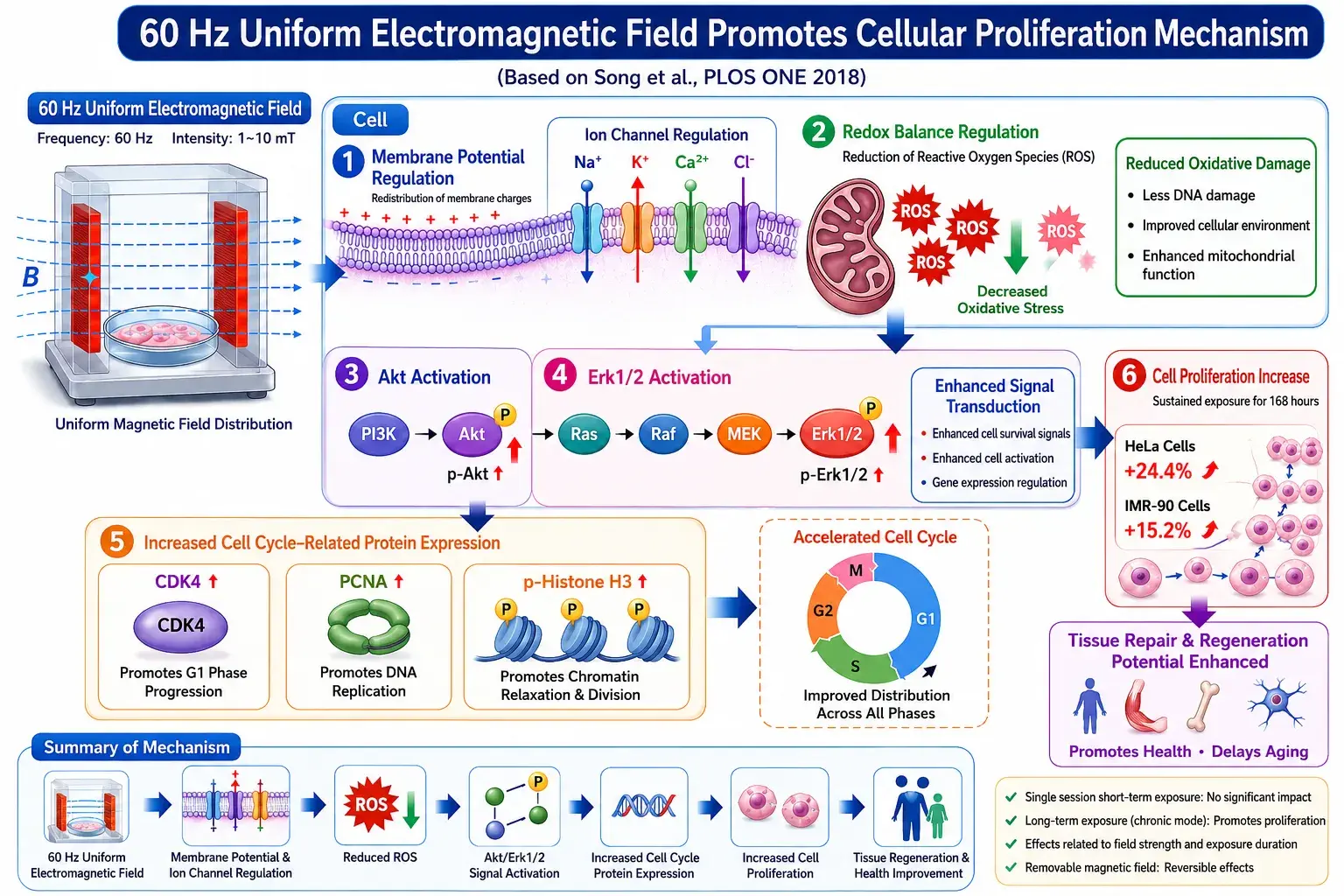
According to research by Song K. et al. published in PLOS ONE (2018), a uniform extremely low frequency electromagnetic field (ELF-EMF) at 60 Hz and 6 mT, under prolonged continuous exposure, does not cause DNA damage or cytotoxicity. Instead, it promotes the proliferation of both normal and cancer cells. The study indicates that this effect is related to a decrease in intracellular reactive oxygen species (ROS) and the activation of cell signaling pathways.
Mechanism of action process
① Uniform electromagnetic field acts on the cell membrane
After a uniform electromagnetic field of 60 Hz acts on cells, it can affect the charge distribution on the cell membrane surface and ion channel activity, thereby improving the efficiency of ion exchange inside and outside the cells, including:
- Sodium ion (Na⁺)
- Potassium ion (K⁺)
- Calcium ion (Ca²⁺)
- Chloride ion (Cl⁻)
This process helps maintain stable membrane potential and efficient cell signaling.
② Reduce intracellular reactive oxygen species (ROS)
Research finds:
After continuous exposure to a 60 Hz uniform electromagnetic field,
Intracellular reactive oxygen species (ROS) levels decrease.
The results include:
- Reduced oxidative stress
- Cellular environment improvement
- Mitochondrial function optimization
- Cellular metabolic efficiency improvement
Research has confirmed that the reduction of ROS is one of the key factors promoting cell proliferation.
3. Aktand Erk1/2 Signaling Pathway Activation
When ROS declines, two important growth signaling pathways within the cell are activated:
AktPath
- PI3K activation
- Akt phosphorylation increased (p-Akt ↑)
- Promote cell survival and growth
ERK1/2Path
- Ras → Raf → MEK → Erk1/2
- Erk1/2 phosphorylation increased (p-Erk1/2 ↑)
- Promote cell growth and gene expression
Research shows:
Both Akt and Erk1/2 phosphorylation levels were significantly increased.
④ Cell cycle protein increase
Following signal activation, the expression of cell cycle-related proteins increases:
CDK4 up
Accelerate G1 phase progression
PCNA increased
Promote DNA replication
Increased p-Histone H3
Promote chromosome condensation and cell division
The cell has entered a more active proliferative state.
⑤ Cell cycle acceleration
Research finds:
Although the proportions of each cell cycle (G1, S, G2, M) did not significantly change,
But the overall cell cycle operates faster.
Representative
- Increased cell division efficiency
- Increased cell turnover rate
- Enhanced proliferative capacity
Rather than remaining in a specific cyclical phase.
⑥ Increased cell proliferation
After 168 hours of continuous exposure:
HeLacell
Proliferation rate increase
+24.4%
IMR-90Fibroblast
Proliferation rate increase
+15.2%
Research also confirmed:
- No DNA damage
- No obvious cytotoxicity
- The effect is positively correlated with exposure time and magnetic field strength.
However, after stopping the exposure, this promoting effect gradually disappears, which is a reversible reaction.
Bioelectric Layered Integration Model
From an electrobiological perspective, it can be explained as:
Membrane Potential
Ion channel regulation
Cell Potential
Signal transduction activation
Body Potential
Multicellular cooperation
ROSDescent
↓
Akt / Erk1/2Activation
↓
Cell cycle protein increase
↓
Increased cell proliferation
↓
Enhanced potential for tissue repair and regeneration
Conclusion
This study shows60 Hz uniform electromagnetic fieldIts main mechanism of action is not direct stimulation of cell division, but rather by regulating cell membrane ion channels and redox balance, reducing intracellular ROS concentration, thereby activating Akt and Erk1/2 signaling pathways, increasing the expression of cell cycle proteins such as CDK4, PCNA, and p-Histone H3, and ultimately promoting cell proliferation and tissue repair potential.Research also confirmed that under the experimental conditions, no DNA damage or cytotoxicity was observed.